Cell engineering:
Progress in the retinal stem cell field is hampered by a lack of cell based reporter tools to monitor development. Towards this goal we are developing gene-editing tools to tag fluorescent reporters to genes that are expressed in the retina. These tools will allow us to monitor the presence / absence of photoreceptor and ganglion cells which will in turn facilitate studies of eye development and disease.
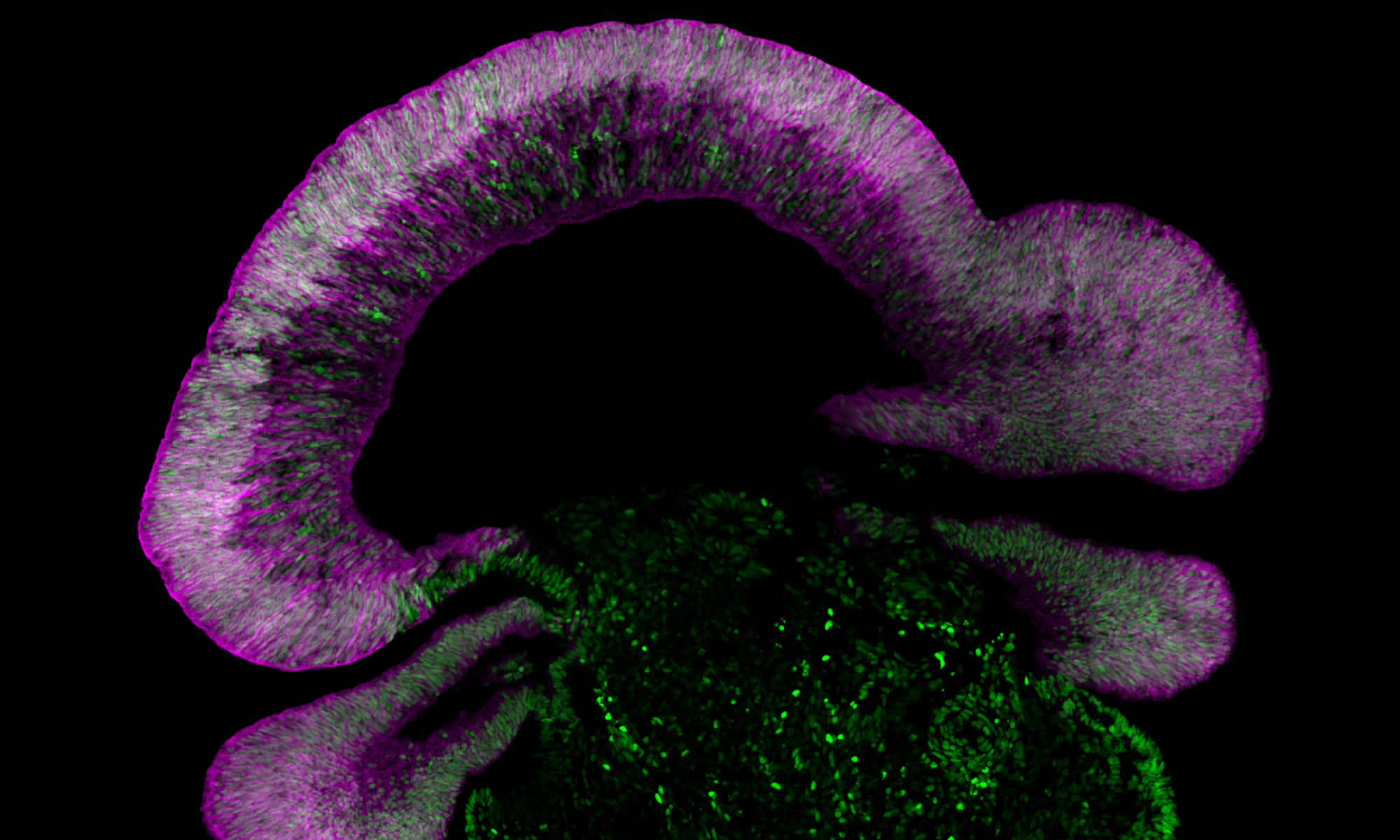
Retinal development:
Human retinal organoids resembling actual human retinas offer a convenient way to study human eye development in vitro. Our lab uses retinal reporters to monitor the development of human photoreceptors and ganglion cells.
Small molecule drug screening:
We are actively screening small molecule drugs to identify factors that increase the generation of retinal cells in 3D “mini-retinas” and 2D dissociated monolayer cultures.
Retinal disease modeling:
Using genetically matched CRISPR-Cas9 gene edited cells. We are currently developing models for retinal degeneration by targeting genes known to be involved in retinal degeneration. To make retinal disease models more accurate we are investigating how microglia influence neuronal disease progression. While this has direct applications in retinal degenerations, microglia may also have possible applications in the eye during Alzheimers disease.
In vivo regeneration (endogenous regeneration) :
Many species, including amphibians, fish and birds, are capable of remarkable levels of repair. Unfortunately, this repair capability seems to have been lost, or dramatically reduced, in higher mammals. A holy-grail of vision restoration is to tap into this endogenous repair in human cells. Our lab is exploring possible strategies to do this using human stem cell derived 3D retinas grown in vitro. Potential applications for this exciting work include restoration of rod and cone photoreceptors in Leber congenital amaurosis/Retinitis pigments, foveal hypoplasia, and glaucoma.