Members of the Toomey lab attending The San Diego Glycobiology Symposium (SDGS), where are research volunteers Kamalu Park and Madeline Mozafari presented posters at the event.
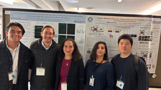
Left to right; Kamalu Park (Undergraduate Research Volunteer, Team leader, Dr. Christopher Toomey, Madeline Mozafari, Research Volunteer, Sima Parvizi Omran, Graduate Student and Jaesoo Jung, Postdoctoral Scholar).